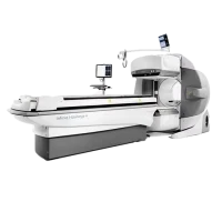
GE Infinia Hawkeye SPECT Gamma Camera
The GE Infinia Hawkeye is used for obtaining detailed images of the body's internal structures by detecting gamma rays emitted from radioactive tracers administered to the patient. It is particularly valuable for cardiac imaging, oncology, and evaluating various physiological functions.
Key Features:
1. SPECT Imaging:
- Technology: Utilizes SPECT imaging technology to provide three-dimensional images of radiotracer distribution within the body. This allows for precise localization of abnormalities and detailed functional information.
- High Sensitivity: Equipped with high-sensitivity detectors to enhance image quality and improve the detection of low levels of radioactivity.
2. Hybrid Imaging:
- Hawkeye Technology: The "Hawkeye" designation indicates that the system integrates SPECT with CT (Computed Tomography) capabilities. This hybrid approach provides anatomical localization and helps correlate functional imaging data with structural images.
- Fusion Imaging: Allows for fusion of SPECT and CT images to enhance diagnostic accuracy, particularly in oncology and cardiac imaging.
3. Advanced Detection:
- Detector Design: Features advanced detector technology to capture gamma rays more efficiently, improving image resolution and contrast.
- Collimator Options: Includes various collimators (e.g., low-energy high-resolution or high-energy) to optimize imaging based on specific diagnostic needs.
4. User Interface:
- Control and Analysis Software: Comes with sophisticated software for controlling the system, processing images, and analyzing data. The software typically includes tools for image reconstruction, quantification, and reporting.
- Ergonomics: Designed for ease of use with a user-friendly interface and options for customizing imaging protocols.
5. Workflow Efficiency:
- Automated Features: Offers automated features to streamline imaging procedures, reduce manual labour, and improve throughput.
- Data Management: Integrates with hospital information systems (HIS) and radiology information systems (RIS) for efficient data management and image storage.
Clinical Applications:
1. Cardiology:
- Myocardial Perfusion Imaging: Assesses blood flow to the heart muscle and identifies areas with reduced or compromised perfusion, useful for diagnosing coronary artery disease.
- Stress Testing: Can be used in conjunction with stress testing to evaluate heart function under physical or pharmacologic stress.
2. Oncology:
- Tumor Detection: Helps in detecting and monitoring tumours and metastatic disease by evaluating the distribution of radiotracers in cancerous tissues.
3. Neurology:
- Brain Imaging: Useful for imaging brain function and detecting abnormalities in conditions like epilepsy and dementia.
