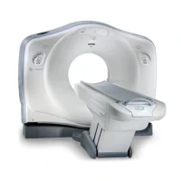
GE VCT 64 Slice CT Scan Machine
The GE VCT 64 Slice CT Scan Machine, also known as the GE VCT (Volume Computed Tomography) 64, is a high-performance computed tomography (CT) scanner designed for advanced imaging with enhanced resolution and speed. Here’s an overview of its key features, operation, and maintenance:
Key Features:
1. 64-Slice Technology:
- High Resolution: Offers 64 slices per rotation, which provides high-resolution images and improved anatomical detail.
- Fast Imaging: Capable of capturing detailed images quickly, which is beneficial for imaging dynamic processes and reducing scan times.
2. Advanced Imaging Capabilities:
- Volume Scanning: Provides high-quality volumetric imaging for comprehensive diagnostic information.
- Multi-Detector Array: Utilizes multiple detector rows to improve image quality and coverage.
3. Enhanced Patient Safety and Comfort:
- Reduced Scan Time: Faster imaging reduces the time patients need to spend in the scanner.
- Low Radiation Dose: Features dose reduction technologies to minimize radiation exposure to patients.
4. User-Friendly Interface:
- Control Console: Equipped with an advanced control console for easy setup, operation, and image review.
- Advanced Software: Comes with software for image reconstruction, post-processing, and data analysis.
5. Versatility:
- Diagnostic Applications: Suitable for a wide range of applications, including cardiology, oncology, and emergency imaging.
Operation:
1. Patient Preparation:
- Positioning: Position the patient on the scanner table according to the imaging protocol. Ensure the patient is comfortable and properly aligned.
- Contrast Administration: If required, administer contrast agents as per the protocol for enhanced imaging of specific tissues or organs.
2. Scan Setup:
- Select Protocol: Choose the appropriate scanning protocol from the control console or software interface. Adjust parameters such as slice thickness, scan range, and reconstruction settings.
- Initialize Scan: Start the scan. The machine will rotate around the patient, capturing multiple slices of data.
3. Image Acquisition:
- Real-Time Monitoring: Monitor the scan process and adjust settings as necessary to ensure optimal image quality.
- Post-Processing: Use the software tools to reconstruct, analyze, and review the images.
4. Data Management:
- Save and Review: Save the images and data. Review them using the diagnostic software and prepare reports as needed.
