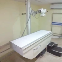
GE DX525 X-Ray Machine
The GE DX525 X-Ray Machine is a digital radiography system designed for high-quality imaging with advanced features to support various diagnostic needs. Here’s a comprehensive overview of its key features, operation, maintenance, and troubleshooting tips:
Key Features:
1. Digital Radiography:
- High-Resolution Imaging: Provides high-quality digital images with improved clarity and contrast.
- Efficient Workflow: Reduces processing time and enhances workflow efficiency with digital image acquisition.
2. Advanced Technology:
- Flat Panel Detector: Uses a flat panel detector to capture X-ray images, offering superior image quality and quick image acquisition.
- Automatic Exposure Control: Adjusts exposure settings automatically based on the patient’s size and the imaging area to optimize image quality and reduce radiation dose.
3. Versatility:
- Multi-Modal Capabilities: Suitable for a range of applications, including general radiography, fluoroscopy, and specialized imaging procedures.
- Flexible Positioning: Designed for ease of use with adjustable table height and imaging angles to accommodate various patient positions and needs.
4. User-Friendly Interface:
- Control Console: Features an intuitive control console for easy operation, image acquisition, and workflow management.
- Advanced Software: Includes software for image processing, analysis, and storage, with options for integration into hospital information systems (HIS) or picture archiving and communication systems (PACS).
5. Patient Safety:
- Dose Optimization: Incorporates dose-reduction technologies to minimize radiation exposure while maintaining diagnostic image quality.
- Ergonomic Design: Designed with patient comfort in mind, including features to ease patient positioning and reduce discomfort during imaging.
Operation:
1. Patient Preparation:
- Positioning: Position the patient correctly on the examination table based on the required imaging procedure. Ensure proper alignment and comfort.
- Shielding: Use appropriate lead shielding to protect sensitive areas from unnecessary radiation.
2. Image Acquisition:
- Select Protocol: Choose the imaging protocol from the control console or software interface based on the clinical indication.
- Adjust Settings: Configure exposure parameters, including technique factors, to suit the patient and the area of interest.
- Capture Image: Initiate the imaging process. The flat panel detector will capture the X-ray image, which is then processed and displayed digitally.
3. Image Review and Management:
- Analyze Images: Review the images on the control console or connected workstation. Use software tools for image enhancement, measurements, and annotations.
- Save and Transfer: Save images and patient data. Transfer images to PACS or other storage systems as needed.
Maintenance Tips:
1. Regular Cleaning:
- Detector and Table: Clean the flat panel detector and examination table regularly to maintain hygiene and image quality.
- X-Ray Tube: Keep the X-ray tube and surrounding area clean to ensure optimal performance.
2. Calibration:
- Routine Calibration: Perform regular calibration and quality control checks to ensure accurate image acquisition and consistent performance.
- Software Updates: Update the imaging software to the latest version for improved functionality and performance.
3. Technical Support:
- Service Agreements: Consider a service agreement with GE Healthcare for scheduled maintenance and technical support.
- Troubleshooting: Refer to the user manual for troubleshooting guidance or contact technical support for assistance with operational issues.
4. Preventive Maintenance:
- Scheduled Maintenance: Follow the manufacturer’s recommended maintenance schedule to ensure the longevity and reliability of the machine.
