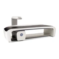
GE Lunar Bone DEXA Machine
The GE Lunar Bone DEXA (Dual-Energy X-ray Absorptiometry) Machine is a diagnostic tool used for measuring bone mineral density (BMD) to assess bone health and diagnose conditions such as osteoporosis. It provides precise and reliable measurements by using X-ray technology. Here’s an overview of its key features, operation, maintenance, and troubleshooting tips:
Key Features:
1. High-Precision Imaging:
- Dual-Energy X-ray Absorptiometry: Utilizes two different X-ray energy levels to differentiate between bone and soft tissue, allowing for accurate measurement of bone mineral density.
- High Resolution: Provides high-resolution images to ensure precise bone density measurements.
2. Advanced Technology:
- GE Lunar Technology: Features advanced GE Lunar technology, known for its accuracy and reliability in BMD assessment.
- Automated Analysis: Includes software for automated analysis and reporting, which helps in quick and accurate interpretation of results.
3. Versatility:
- Multiple Scan Sites: Capable of scanning various sites such as the lumbar spine, hip, and forearm, providing a comprehensive assessment of bone health.
- Low Radiation Dose: Designed to deliver minimal radiation exposure to patients, ensuring safety while maintaining diagnostic accuracy.
4. User-Friendly Interface:
- Control Console: Features an intuitive control console for easy setup, operation, and data management.
- Software Integration: Includes software for image acquisition, analysis, and integration with electronic medical records (EMR) or hospital information systems (HIS).
5. Patient Comfort:
- Ergonomic Design: Designed with patient comfort in mind, including features to facilitate easy positioning and minimize discomfort during scanning.
Operation:
1. Patient Preparation:
- Positioning: Position the patient on the scanning table according to the required scan site (e.g., lumbar spine, hip). Ensure the patient is properly aligned and comfortable.
- Clothing and Accessories: Remove any metal objects, jewelry, or clothing that could interfere with the scan.
2. Scan Setup:
- Select Protocol: Choose the appropriate scan protocol from the control console or software interface based on the clinical indication and the area to be scanned.
- Adjust Settings: Configure scan parameters, such as scan region and resolution, as needed.
3. Image Acquisition:
- Initiate Scan: Start the scanning process. The machine will use dual-energy X-ray beams to measure bone density at the selected site.
- Monitor Scan: Ensure the scan is completed without any issues or artifacts.
4. Data Analysis and Reporting:
- Review Images: Use the software to review and analyze the images. Generate reports based on the BMD measurements and any other relevant findings.
- Save and Transfer: Save the images and reports. Transfer the data to EMR or HIS as required.
