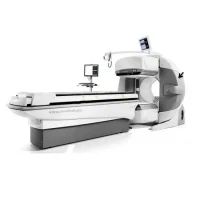
Single Photon Emission Computed Tomography (SPECT) scan
SPEC CT typically refers to a type of imaging technology that combines Single Photon Emission Computed Tomography (SPECT) with Computed Tomography (CT). This hybrid imaging modality is designed to provide both functional and anatomical information, offering enhanced diagnostic capabilities for various medical conditions.
Key Features:
1. Hybrid Imaging:
- SPECT Imaging: SPECT is a nuclear medicine technique that provides functional imaging by detecting gamma rays emitted from a radiotracer injected into the patient. It is used to assess physiological processes and abnormalities.
- CT Imaging: CT provides precise anatomical pictures utilising X-rays. It helps in visualizing the precise location of abnormalities detected by SPECT.
2. Enhanced Diagnostic Capabilities:
- Functional and Anatomical Information: The combination of SPECT and CT allows for the fusion of functional data (from SPECT) with anatomical data (from CT), improving diagnostic accuracy.
- Localization: Helps in accurately localizing and characterizing abnormalities, which is crucial for treatment planning and follow-up.
3. Image Fusion:
- Data Integration: The system integrates SPECT and CT images to create a single dataset where functional and anatomical information are aligned, enhancing the ability to diagnose and assess medical conditions.
4. Improved Accuracy:
- Precise Localization: Provides precise localization of functional abnormalities within anatomical structures, leading to better diagnosis and treatment planning.
- Enhanced Resolution: Combines the high resolution of CT with the functional imaging capabilities of SPECT.
5. Clinical Applications:
- Cardiology: Used for assessing myocardial perfusion, detecting coronary artery disease, and evaluating cardiac function.
- Oncology: Helps in detecting and staging tumours, assessing response to therapy, and monitoring for recurrence.
- Neurology: Assists in evaluating brain function, diagnosing and monitoring neurological disorders, and assessing cerebral blood flow.
6. Patient Preparation and Safety:
- Radiotracer Administration: A radiotracer is administered to the patient, which is typically done through intravenous injection. The choice of radiotracer depends on the clinical indication.
- CT Scanning: The CT component uses X-ray technology, so the patient is exposed to a small amount of radiation, but the benefits often outweigh the risks in diagnostic contexts.
Advantages:
1. Comprehensive Imaging: Provides both functional and anatomical insights in a single imaging session, offering a more complete picture of the patient’s condition.
2. Improved Diagnosis: Enhances diagnostic accuracy by allowing clinicians to correlate functional abnormalities with precise anatomical locations.
3. Efficient Workflow: Streamlines the imaging process by combining two modalities into one system, potentially reducing the need for separate imaging sessions.
