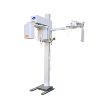
OPG (DENTAL X-RAY) Machine
An Orthopantomogram (OPG) machine, also known as a panoramic dental X-ray machine, provides a comprehensive view of the entire mouth in a single image. It captures a broad and detailed image of the teeth, jaws, and surrounding structures, which is useful for a variety of diagnostic and treatment planning purposes. Here’s a more detailed look at its features, operation, and applications:
Key Features:
1. Panoramic Imaging
- Wide Coverage The OPG machine takes a single, continuous image that covers the entire dental arch, including both upper and lower jaws, and the surrounding structures.
- Rotating Mechanism The X-ray tube rotates around the patient’s head to capture the panoramic view.
2. Digital Technology
- Digital Sensors Modern OPG machines use digital sensors or plates instead of traditional film. This provides instant image acquisition, higher resolution, and the ability to digitally enhance and manipulate the images.
- Image Processing Software Digital systems come with software that allows for image adjustment, contrast enhancement, and detailed analysis.
3. Patient Positioning
- Head Supports The machine includes head supports and alignment guides to ensure proper positioning of the patient’s head during the X-ray. Proper positioning is crucial for accurate imaging and minimizing artefacts.
4. Radiation Safety
- Low Dose The OPG machine is designed to minimize radiation exposure to the patient. Safety features include collimation to focus the X-ray beam and lead aprons to protect other parts of the body.
- Automatic Exposure Control Some models feature automatic exposure control to adjust the radiation dose based on the patient’s size and the area being imaged.
Operation:
1. Preparation
- Patient Instructions The patient is instructed to remove any metal objects, such as earrings and dentures, and to stand still during the imaging process.
- Positioning The patient’s head is positioned using the provided supports, and they are instructed to bite down on a bite block or positioning device to ensure accurate alignment.
2. Imaging Process
- X-Ray Capture The machine’s X-ray tube rotates around the patient’s head while the detector captures the X-ray images. The panoramic image is created by combining multiple X-ray images into a single view.
- Exposure Time The actual exposure time is relatively short, typically lasting only a few seconds.
3. Image Review
- Immediate Results Digital systems provide immediate results, allowing the dental professional to review the images on a computer screen.
- Analysis The images can be analyzed using the accompanying software, which may include tools for measuring bone density, detecting abnormalities, and planning treatments.
Applications:
1. Diagnostic Evaluation
- Comprehensive View Provides a detailed overview of dental and bone structures, aiding in the diagnosis of conditions such as cavities, bone loss, and impacted teeth.
- Pathology Detection Useful for identifying cysts, tumors, and other abnormalities in the jaw and surrounding tissues.
2. Treatment Planning
- Orthodontics Helps in planning orthodontic treatments by showing the alignment of the teeth and the relationship between the upper and lower jaws.
- Implant Planning Assists in evaluating the bone structure and density for the placement of dental implants.
3. Monitoring and Follow-Up
- Progress Tracking Useful for monitoring changes over time, assessing the progress of treatments, and planning further interventions if needed.
Advantages:
1. Broad Coverage Captures a wide area in a single image, reducing the need for multiple X-rays and providing a comprehensive view.
2. Efficient The imaging process is quick, and digital technology allows for immediate viewing and processing of images.
3. Low Radiation Dose Modern OPG machines are designed to minimize radiation exposure to the patient.
