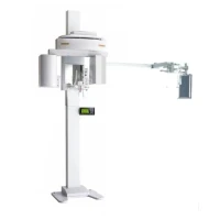
Apex OPG X-Ray System
The Apex OPG X-ray System is a type of panoramic dental X-ray machine designed to capture a comprehensive image of the entire dental arch and surrounding structures. It is used for diagnosing and planning treatments related to dental and jaw conditions.
Key Features:
1. Panoramic Imaging:
- Wide Coverage: Provides a broad view of both the upper and lower jaws, including the teeth, bone structures, and surrounding tissues in a single image. This panoramic view is useful for diagnosing a variety of dental conditions.
2. Advanced Imaging Technology:
- Digital Sensors: Often equipped with digital sensors that capture high-resolution images, offering better image quality and immediate results compared to traditional film-based systems.
- Automatic Exposure Control: Includes features to automatically adjust the X-ray exposure based on the patient’s size and the area being imaged, optimizing image quality and minimizing radiation exposure.
3. User-Friendly Interface:
- Control Panel: Features an intuitive control panel or touchscreen interface for easy operation and programming of imaging settings.
- Image Processing Software: Comes with advanced software for image acquisition, processing, and analysis. This software often includes tools for enhancing image contrast, zooming, and measuring anatomical structures.
4. Patient Positioning:
- Head Supports and Guides: Equipped with adjustable head supports and alignment guides to ensure accurate positioning of the patient during the imaging process. Proper positioning is crucial for obtaining high-quality images and minimizing artifacts.
5. Radiation Safety:
- Minimized Exposure: Designed with safety features to limit radiation exposure to the patient. This includes collimation to focus the X-ray beam and lead aprons to protect other parts of the body.
- Regulated Dose: Ensures that the radiation dose is kept as low as reasonably achievable while still providing clear images.
Operation:
1. Preparation:
- Patient Instructions: The patient is asked to remove any metal objects, such as earrings and dentures, and to wear a lead apron for protection.
- Positioning: The patient’s head is positioned using the provided supports, and they are instructed to bite down on a positioning device to ensure proper alignment.
2. Imaging Process:
- X-Ray Capture: The X-ray tube rotates around the patient’s head, capturing a panoramic image as the tube moves in a horizontal arc.
- Exposure Time: The exposure time is relatively short, usually only a few seconds, during which the patient needs to remain still.
3. Image Review:
- Immediate Results: Digital systems provide immediate images that can be reviewed on a computer screen. The images can be analyzed and adjusted using the accompanying software.
Applications:
1. Diagnostic Evaluation:
- Comprehensive View: Used to assess the overall condition of the teeth, bone structures, and surrounding tissues, helping in diagnosing conditions such as cavities, bone loss, and impacted teeth.
- Pathology Detection: Useful for identifying abnormalities such as cysts, tumours, and other pathological conditions in the jaw and surrounding areas.
2. Treatment Planning:
- Orthodontics: Helps in planning orthodontic treatments by providing a complete view of dental alignment and the relationship between the upper and lower jaws.
- Implant Planning: Assists in evaluating bone structure and density for the placement of dental implants.
3. Monitoring:
- Progress Tracking: Allows for monitoring changes in dental and bone structures over time, aiding in the assessment of treatment progress or disease progression.
Advantages:
1. Broad Coverage: Provides a wide-area view in a single image, reducing the need for multiple X-ray images and offering a comprehensive assessment.
2. Efficiency: The imaging process is quick, and digital systems provide immediate results, improving patient throughput and convenience.
3. Enhanced Image Quality: Digital technology allows for high-resolution images with the ability to enhance and manipulate the images for better diagnosis.
